Persistent Hydration Sites and Water Exchange in Daraxonrasib Simulations
by Ishaan Ganti · October 19, 2026
Muir Glacier, Alaska by Thomas Hill.
A while ago, I ran some molecular dynamics (MD) simulations of the daraxonrasib-KRAS G12V-CypA complex (using a prepared version of 9BG6 in the PDB). Unsurprisingly, the ligand stayed close to its starting pose in every replica I ran. But some cool things emerged from our trajectory post-processing, specifically pertaining to hydration sites! We've previously explored hydration sites and their role in protein–ligand interactions. Rowan reports both how often each site is occupied and how many distinct waters visit it. A site can stay occupied even as individual water molecules come and go.
Rowan's hydration-site analysis revealed around 10 high-occupancy hydration sites around the protein–ligand interface, of which two features were particularly interesting.
The first was an extremely persistent water bridge between daraxonrasib and KRAS. In one pair of 50 ns replicas, the hydration site was occupied in 95% of frames and formed a protein–ligand bridge in 92% of frames. I reran the system without hydrogen mass repartitioning (HMR), to make sure that results persisted with more realistic water dynamics, and got essentially the same result: 96% occupancy and an 84% bridging frequency. Interestingly, these weren't singular water molecules that happened to get trapped; in the two runs, 17 and 28 distinct waters visited the site. A bridging site featuring this sort of exchange isn't something I've seen much before; this indicates that the site is thermodynamically favored but water exchange is kinetically facile.
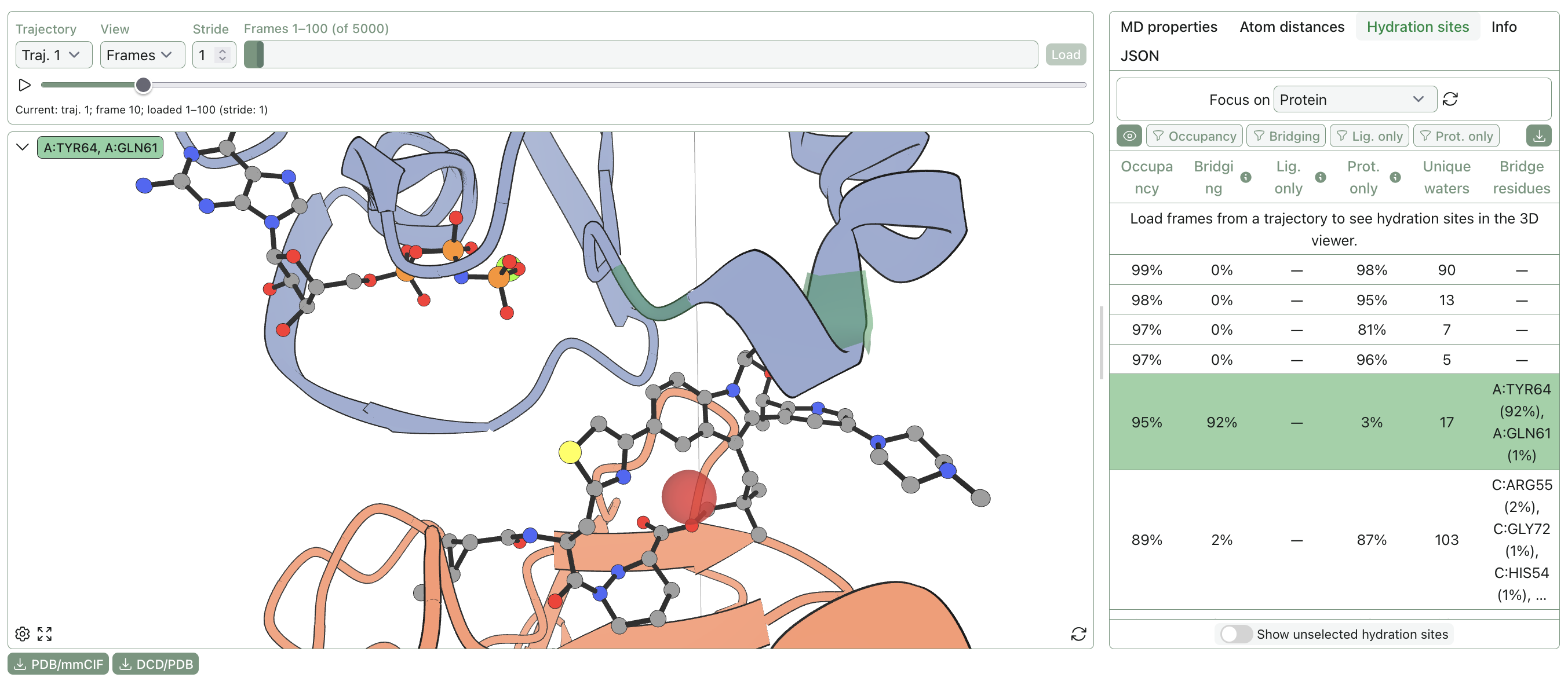
The bridging hydration-site at the binding interface. The hydration-site is depicted as a translucent red sphere.
This lines up nicely with the experimental structure. Daraxonrasib's thiazole has been reported to form a water-bridged hydrogen bond with KRAS Tyr64 in PDB 9BG6. We think it's cool that pretty standard explicit-solvent MD recovers a water-mediated interaction independently identified from crystallography (the solvent was stripped and re-added before simulation, so the crystallographic waters were not retained).
The second feature I noticed was that several high-occupancy sites form a spatial cluster extending from daraxonrasib to GNP, the non-hydrolyzable GTP analog present in 9BG6. Some neighboring hydration-site centroids are only a few Ångstroms apart, geometrically compatible with a connected hydrogen-bonded water network. And, this broader hydration pattern is pretty reproducible: across both simulations (HMR and non-HMR), the strongest sites appear in roughly the same positions.
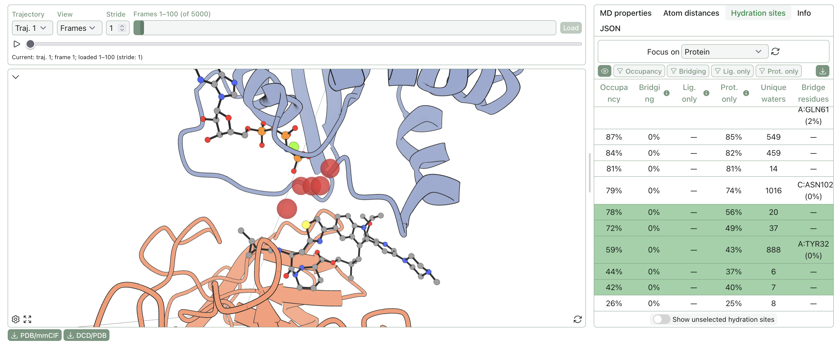
Hydration-sites in the gap between GNP and daraxonrasib in the MD simulation without HMR. The leftmost site is the one with the highest (highlighted in green in the table) exchange of the five.
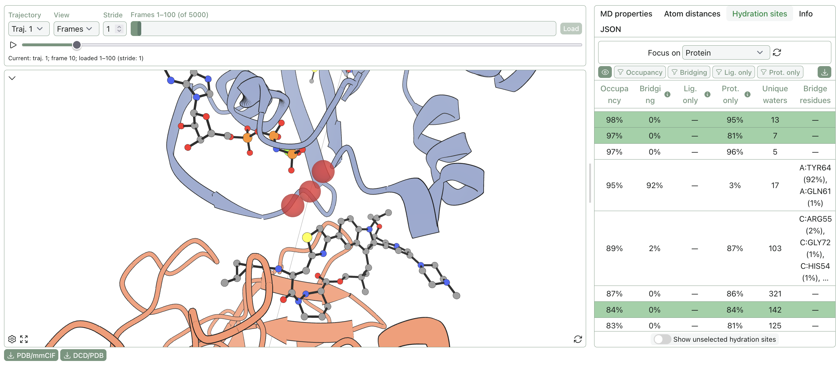
Hydration-sites in the gap between GNP and daraxonrasib in the MD simulation with HMR. The leftmost site is the lowest occupancy of the three and has the highest (highlighted in green in the table) exchange of the three.
The unique water counts are, once again, worth paying attention to. All of these sites have relatively low unique water counts, with some being single-digit, except for a single site in each of the MD simulations. In both of the above images, they are the leftmost sites. As such, these are likely more exposed to the bulk solvent. This made me think that the nearby protein residues might be "trapping" some waters in a small pocket. Turning on the residue atoms nearby lets us inspect this visually!
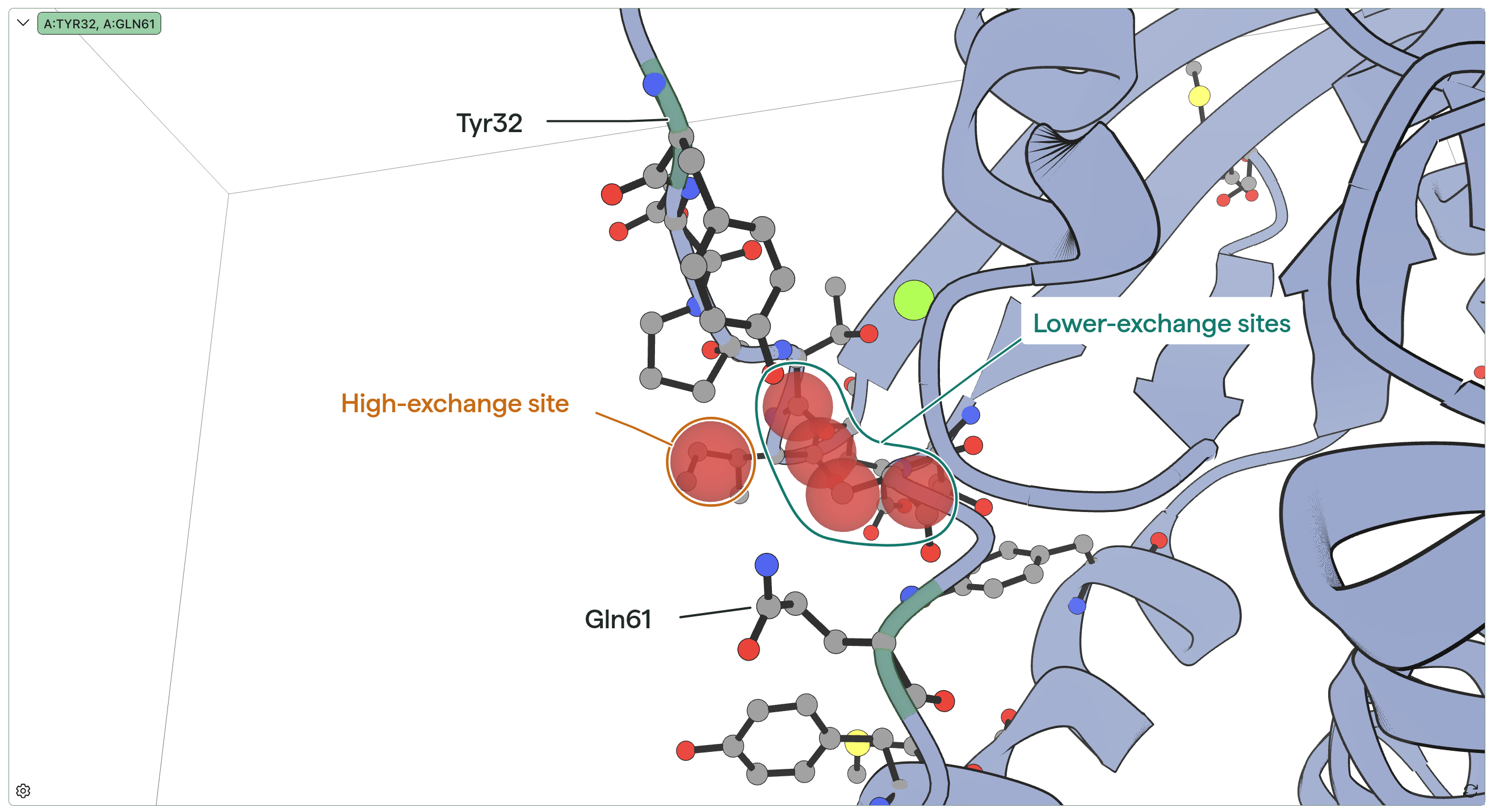
The Tyr32 and Gln61 residues loosely seem to separate the very high-exchange hydration-site from the other sites. The other protein chain and daraxonrasib are hidden for viewing ease.
It generally looks like Tyr32 and Gln61 create a small barrier between the high-exchange site and the low-exchange sites inside this little divot. And, cool enough, work on closely related systems has discussed water networks being formed through interactions with Tyr32! For example, see Fig. 1c here.
These two features illustrate how looking at conserved hydration sites can provide interesting and non-obvious structural insights. Water networks and hydration sites adapt in complex and dynamic ways to different protein–ligand structures; if you're looking to study hydration sites in your complexes, try Rowan out!
See the calculations: